
EPINOV: A Computer Model of Epileptic Brain Undergoes Clinical Trial for Transforming Surgery
April 11, 2023
Article published by NewsClick
In the clinical trial named EPINOV, scientists of the Institute De Neuroscience Des Systems (INS) in France are looking at how far a computer model of the epileptic brain can improve the rate of epilepsy surgeries. They have reported on the approach and the clinical trial in their research paper published in the journal Science.
The models are created using the Virtual Epileptic Patient (VEP), which is a computational system developed by scientists. This project is a part of the Human Brain Project (HBP), a 10-year research project started in 2013 with an aim of allowing researchers across Europe to advance their understanding of neuroscience and brain diseases.
In an article by Miryam Naddaf published at Nature Briefing, UCL (University College London) neurosurgeon Aswin Chari, commenting on VEP was quoted as saying, “It’s an example of personalized medicine. VEP uses “the patient’s own brain scans [and] the patient’s own brainwave-recording data to build a model and improve our understanding of where their seizures are coming from.”
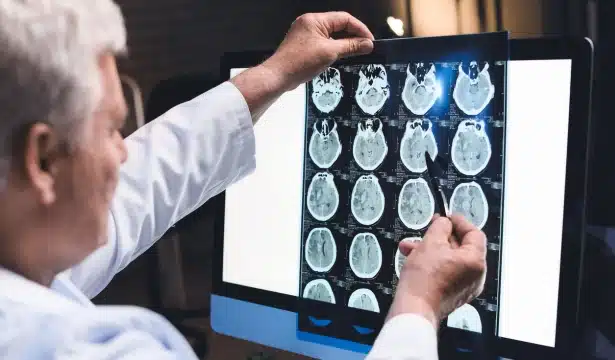
Nearly 50 million people that suffer from epilepsy across the globe, have abnormal brain activities and one-third of them do not respond to drugs applied for stopping epileptic seizures. According to Chari, surgeries are huge reliefs for these patients. In such surgeries, a portion of the brain known as the epileptogenic zone, which initiates the seizures, are removed. It is still a challenge for neurosurgeons to find out the exact portion of the epileptogenic zone. Remember, brain surgeries always come with challenges, especially in finding the exact location a surgeon will make the incision. A cut in an unwanted portion can lead to severe conditions like paralysis.
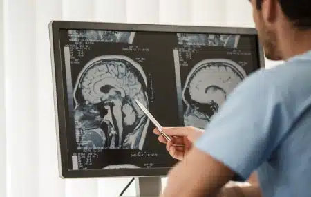
In epileptic surgeries, clinicians take advantage of several techniques, especially to find the epileptogenic zone, such as MRI (Magnetic Resonance Imaging), EEG (Electroencephalogram) etc. Surgeons also use the SEEG (Stereoelectroencephalography) technique. Here, seven centimetres-long 16 electrodes are placed over the skull and the electrodes record the brain waves (depicting brain activities) of specific areas for a week or two.
Latest from the Community