
Experts Use MRI to Pinpoint Link Between Pediatric TBI and Cognitive Impairment
July 14, 2022
Article published by Health Imaging
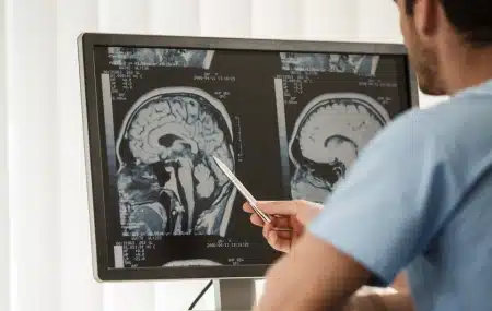
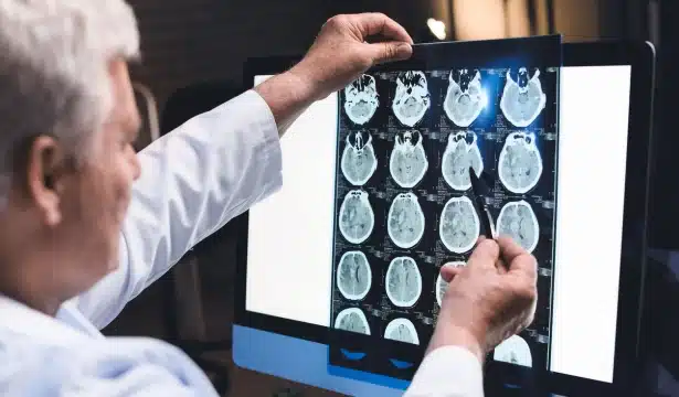
Experts recently found reduced brain volumes in pediatric patients who had previously sustained serious head injuries.
New work published in Brain details how researchers concluded that both gray and white matter volumes decrease relative to head size during adolescent development. These findings were associated with certain cognitive and emotional impairments—a correlation that has been difficult to conclude in pediatric populations, first author Niall Bourke, from the Department of Brain Sciences at Imperial College London, and colleagues shared:
“In adults, traumatic brain injury [TBI, which can lead to the development of a type of epilepsy known as post-traumatic epilepsy] produces progressive brain atrophy that can be accurately measured and is associated with cognitive decline. However, the effect of pediatric traumatic brain injury on brain volumes is more challenging to measure because of its interaction with normal brain development.”
TBI patients displayed reductions of both gray and white matter on imaging in comparison to their peers. At least one white matter tract with reduced volume—most often in midline white matter structures including the corpus callosum—was observed in 28% of TBI patients, and 18% displayed similar findings in gray matter tracts.
Those with lower brain volumes were also reported to have slower processing speeds, emotional impairment, learning difficulties, increased apathy and anger.
“These individualized assessments of volume can provide some understanding of related cognitive function following TBI. Alongside other clinical tools, age-specific volume estimates can aid the clinical picture of an injury to better understand the individual case.”
Latest from the Community