
Study: Computer-Assisted Planning for the Insertion of Stereoelectroencephalography Electrodes for the Investigation of Drug-Resistant Focal Epilepsy: an External Validation Study
April 13, 2018
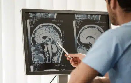
OBJECTIVE: One-third of cases of focal epilepsy are drug refractory, and surgery might provide a cure. Seizure-free outcome after surgery depends on the correct identification and resection of the epileptogenic zone. In patients with no visible abnormality on MRI, or in cases in which presurgical evaluation yields discordant data, invasive stereoelectroencephalography (SEEG) recordings might be necessary. SEEG is a procedure in which multiple electrodes are placed stereotactically in key targets within the brain to record interictal and ictal electrophysiological activity. Correlating this activity with seizure semiology enables identification of the seizure-onset zone and key structures within the ictal network.
The main risk related to electrode placement is hemorrhage, which occurs in 1% of patients who undergo the procedure. Planning safe electrode placement for SEEG requires meticulous adherence to the following: 1) maximize the distance from cerebral vasculature, 2) avoid crossing sulcal pial boundaries (sulci), 3) maximize gray matter sampling, 4) minimize electrode length, 5) drill at an angle orthogonal to the skull, and 6) avoid critical neurological structures.
The authors provide a validation of surgical strategizing and planning with EpiNav, a multimodal platform that enables automated computer-assisted planning (CAP) for electrode placement with user-defined regions of interest.
Latest from the Community