

U.Va. Researchers Develop a New Imaging Approach to Target Epilepsy Seizures
July 3, 2021
Article, published in The Cavalier Daily
Summary
An improved form of positron-emission tomography detects glucose in the brain in an effort to prevent seizures.
U.Va. Health neurologist Mark Quigg, radiology and medical imaging Assist. Prof. Bijoy Kundu and Class of 2020 alumnus Vikram Seshadri launched a small pilot study using seven participants to test out a new technique to improve how diagnoses are made. Their goal was to refine the method of glucose mapping for more accuracy and better resolution.
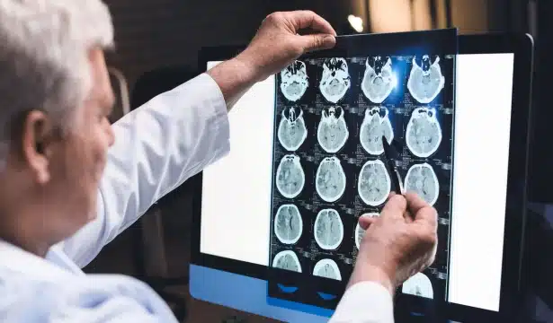
“Normally in a PET scan when we’re taking a picture of glucose use in the brain we use a radioactive tracer,” Quigg said. “What [Kundu] has invented is a process of a dynamic PET.”
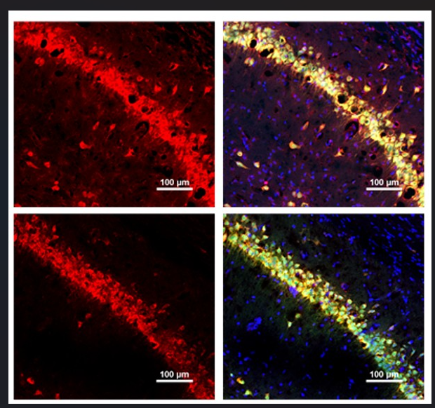
In the dynamic PET scan, a tracer is injected into the bloodstream while the patient is already in the scanner. The scanner then takes pictures like a stop motion animation movie. The result is a time map of glucose uptake and glucose use in relation to brain region. The time map is used to identify points in time that yield the most difference among the functional places in the brain, or areas with most glucose uptake, and the nonfunctional places in the brain, or areas with least uptake.
The key difference between the former way of imaging and the dynamic method lies in the way the images are taken. Instead of producing a single snapshot of the brain, the dynamic PET captures a series of snapshots that then require mathematical modeling for image analysis.
This new technique poses a huge advantage because the results are more quantitative and rigid, which allows for clearer data and less subjectivity compared to the previous PET scan method.
Quigg believes that the findings from this research are very promising for new treatment plans. The new diagnostic approach is completely non-invasive and enables more accurate diagnoses.
Latest from the Community