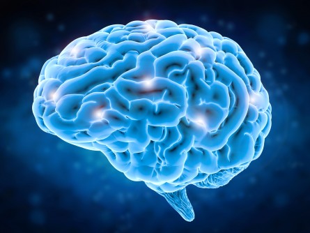

Upper Cebellar Glucose Hypermetabolism in Patients with Temporal Lobe Epilepsy and Interictal Psychosis
August 19, 2022
Abstract found on Wiley Online Library
Objective: Psychosis is an important comorbidity in epilepsy, but its pathophysiology is still unknown. The imaging modality 18F-fluorodeoxyglucose-positron emission tomography (18F-FDG PET) is widely used to measure brain glucose metabolism, and we speculated that 18F-FDG PET may detect characteristic alteration patterns in individuals with temporal lobe epilepsy (TLE) and psychosis.
Methods: We enrolled 13 patients with TLE and interictal psychosis (TLE-P) and 21 patients with TLE without psychosis (TLE-N). All underwent interictal 18F-FDG-PET scanning. Statistical Parametric Mapping (SPM)12 software was used for the normalization process, and we performed a voxel-wise comparison of the TLE-P and TLE-N groups.
Results: Cerebral hypometabolic areas were observed in the ipsilateral temporal pole to hippocampus in both patient groups. In the TLE-P group, the voxel-wise comparison revealed significantly increased 18F-FDG signals in the upper cerebellum, superior cerebellar peduncle, and midbrain. There were no significant between-group metabolic differences around the focus or other cerebral areas.
Significance: Our results demonstrated significant hypermetabolism around the upper cerebellum in patients with temporal lobe epilepsy and interictal psychosis compared to patients with temporal lobe epilepsy without psychosis. These findings may reflect the involvement of the cerebellum in the underlying neurobiology of interictal psychosis and could contribute to a better understanding of this disorder.
Latest from the Community