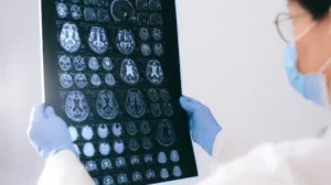
Research Finds Ability to Predict Seizures in Temporal Lobe Epilepsy, 30+ Mins Ahead

Article published by The Mirage

Seizures can be predicted more than 30 minutes before onset in patients with temporal lobe epilepsy, opening the door to a therapy using electrodes that could be activated to prevent seizures from happening, according to new research from UTHealth Houston.

The study, led by Sandipan Pati, MD, associate professor in the Department of Neurology with McGovern Medical School at UTHealth Houston, was recently published in NEJM Evidence, a publication of the New England Journal of Medicine.

“The ability to predict seizures before they occur is a major step forward in the field of epilepsy research,” said Pati, senior author of the study and a member of the Texas Institute for Restorative Neurotechnologies at UTHealth Houston Neurosciences. “These findings are significant because they suggest that we may be able to develop more effective therapies for epilepsy, which could greatly improve the quality of life for patients who suffer from this condition.”

Surgery is a common treatment for many patients with epilepsy. But when seizures affect larger areas of the brain, removing part of the brain surgically is not an option. Neuromodulation therapy could offer an alternative solution for patients with these seizures, Pati said.

Past studies of continuous electroencephalography (EEG) – the measurement and recording of electrical activity in different parts of the brain – have suggested that seizures in people with focal-onset epilepsies tend to occur during periods of heightened risk, represented by pathologic brain activities known as “pro-ictal states.” The EEG-based detection of pro-ictal states is critical to the success of adaptive neuromodulation, with the early detection of seizures allowing electrodes to be applied therapeutically to the brain’s seizure onset zone and thalamus.

To distinguish these pro-ictal states, Pati’s team studied a prospective, consecutive series of 15 patients with temporal lobe epilepsy who underwent limbic thalamic recordings in addition to routine intracranial EEG for seizure localization. In total, they analyzed 1,800 patient hours of continuous EEG.

Epilepsy Research News: December 2022

This issue of Epilepsy Research News includes summaries of articles on:


Increased Seizures After COVID Compared to the Flu

Researchers have found that the risk of seizures or epilepsy following a COVID infection is significantly higher than after being infected with the flu. The team looked at the health records of people who had been infected with COVID and matched them (so that they were similar in characteristics such as age, sex, and medical conditions) with a group of people who had been infected with the flu. The team then compared the incidence of epilepsy and seizures between the two groups over a six-month period following the initial infection. The rate of new cases of epilepsy or seizures was 0.94% in the people who had COVID, compared with 0.6% in those who had the flu. The team indicated that while the overall risk of seizures was very low, people who had COVID were 55% more likely to develop epilepsy or seizures over the next six months than people who had the flu.

Learn More


New Statistical Tool to Understand Seizures

A new study seeks to understand how some people’s seizures change over time in what is known as a seizure ‘cycle’ and understand how certain triggers might increase or decrease seizure risk, perhaps giving people with epilepsy a better idea of how and why their seizures happen, and to better recognize the early warning signs. The study found that aging itself, as well as common triggers, may be contributing factors to how the medical condition affects those prone to seizures. The researchers studied the seizure diaries of more than 1,000 patients ages 2 months to 80 years and developed a new statistical model to explicitly capture the effect of factors that may drive transitions in seizure risk, looking at factors like antiseizure medications, illness, and menstrual cycles. In examining the way seizure cycles vary in people with epilepsy, the researchers found that individuals in older age groups had shorter “calm” stretches between seizures, while younger age groups had longer stretches. This work paves the way for future studies to further examine seizure cycles.

Learn More


Identification of a Possible Molecule to Treat Temporal Lobe Epilepsy (TLE)

Researchers have recently identified and developed a small molecule called D4 with the potential to treat TLE by suppressing neuroinflammation. The findings suggest that D4 strongly suppresses TLE-induced neuroinflammation, curbs TLE seizures, and increases survival rate in an animal model of TLE. D4 works by blocking “hemichannels” in the brain, which are channels that act as pathways for neuroinflammatory molecules. The researchers note that their findings bring forward a possible new pathway for drug development for epilepsy and also highlight the involvement of neuroinflammation in epilepsy.

Learn More


Pinpointing Brain Areas Involved in GLUT1 Deficiency Syndrome Seizures

A small group of brain cells linked to a circuit in the brain is responsible for setting off whole-brain seizures in a rare form of epilepsy affected by blood sugar levels, a new study suggests. This rare genetic disorder is known as GLUT1 deficiency syndrome. Researchers used a combination of electroencephalography (EEG) as well as brain imaging in humans to show that the seizures started from brain areas called the thalamus and somatosensory cortex. When blood sugar levels dipped, abnormal electrical activity in the circuit formed by these areas spread throughout the brain. The researchers also used an animal model of GLUT1 deficiency syndrome to further investigate this circuit and pinpoint the cell types important in causing an imbalance in inhibitory brain activity compared to excitatory brain activity (which can lead to seizures). The researchers suggested these results could point to a mechanism for seizures in GLUT1 deficiency syndrome that might be targeted as a potential treatment for seizures related to GLUT1 Deficiency syndrome.

Learn More

Stereoelectroencephalography-Based Research on the Value of Drug-Resistant Temporal Lobe Epilepsy Auras: A Retrospective Single-Center Study

Abstract found on PubMed

Purpose: To explore the localization value of drug-resistant temporal lobe epilepsy (TLE) aura for preoperative evaluation, based on stereoelectroencephalography (SEEG), and its prognostic value on the surgical outcome.

Methods: The data of patients with drug-resistant TLE who had SEEG electrodes implanted during preoperative evaluation at the First Affiliated Hospital of the University of Science and Technology of China (Hefei, China) were retrospectively analyzed. The patients were divided into aura-positive and aura-negative groups according to the presence of aura in seizures. To explore the clinical features of aura, we evaluated the localizing and prognostic values of aura for the outcome of anterior temporal lobectomy based on SEEG.

Results: Among forty patients, twenty-seven patients were in the aura-positive group and ten (25.0%) patients had multiple auras. The most common TLE aura was abdominal aura [thirteen (34.2%) patients]. The postoperative seizure frequency was significantly reduced in the preoperative aura-positive patients compared to the preoperative aura-negative patients (P = 0.011). Patients with abdominal (P = 0.029) and single (P = 0.036) auras had better surgical prognoses than aura-negative patients. In the preoperative evaluation, aura-positive patients had a better surgical outcome if the laterality of positron emission tomography-computed tomography (PET-CT) hypometabolism was concordant with the epileptogenic focus identified with SEEG (P = 0.031). A good postoperative epileptic outcome in aura-positive patients was observed among those with hippocampal sclerotic medial temporal lobe epilepsy (P = 0.025).

Conclusion: Epileptic aura is valuable for the localization of the epileptogenic focus. Abdominal aura and single aura were good predictors of better surgical outcomes. Among patients with a preoperative diagnosis of hippocampal sclerosis or with laterality of PET-CT hypometabolism concordant with the epileptogenic focus identified using SEEG, those with aura are likely to benefit from surgery.

Neuroscientists Discover a New Drug Candidate for Treating Epilepsy

Article published by Medical Xpress


Temporal lobe epilepsy (TLE) is one of the most common types of epilepsy worldwide. Although symptomatic medications are available, one-third of TLE patients remain unresponsive to current treatment, so new drug targets are critically needed. A research team co-led by a City University of Hong Kong (CityU) neuroscientist has recently identified and developed a new drug candidate with the potential for effectively treating TLE by suppressing neuroinflammation.

A research team co-led by Dr. Geoffrey Lau Chun-yue, Assistant Professor in the CityU Department of Neuroscience, identified a new, small organic molecule called D4, whose effects the team investigated in treating TLE using a mouse model. The findings suggest that D4 strongly suppresses TLE-induced neuroinflammation, curbs TLE seizures, and increases the animal’s survival rate.

“These are very exciting and encouraging results for translational research in epilepsy,” said Dr. Lau. “We have found a very promising new drug candidate for treating epilepsy that works through a new mechanism—blocking connexin hemichannels. Our findings also highlight the important involvement of neuroinflammation in neurological disorders such as epilepsy.”

Changes in Heart Rate During the Peri-Ictal Period in Focal Epilepsy

Abstract found on PubMed

Objective: We explored changes in heart rate during the peri-ictal period in patients with focal epilepsy, and differences in heart rate changes according to epileptic site and side were assessed.

Methods: A total of 198 epileptic seizures in 102 patients with focal epilepsy, who had a definite epileptogenic focus and had undergone surgical treatment, were assessed from 2014 to 2019. Heart rate was measured manually during the peri-ictal period. Change in heart rate and the time it occurred were assessed and compared between different epileptic sites and sides.

Results: Heart rate increased in 177 (89.4%) of 198 seizures. In 82 (44.8%) of 183 seizures, the change in heart rate occurred before seizure onset. The median period of heart rate change was seven seconds (interquartile range: 3–11 seconds) in seizures with heart rate change before seizure onset. The number of seizures with heart rate increase before seizure onset was significantly greater for medial temporal lobe epilepsy compared to lateral temporal lobe epilepsy (p=0.019) and extratemporal lobe epilepsy (p=0.002).

Significance: A change in heart rate prior to seizure onset is more likely to occur in patients with medial temporal lobe epilepsy, compared to those with lateral temporal lobe epilepsy and extratemporal lobe epilepsy. Patients with medial temporal lobe epilepsy may likely benefit from seizure warning and detection devices.

FSU Team Makes Discovery Advancing Epilepsy Research

Article published by Florida State University News

A team of Florida State University College of Medicine researchers has found a link between a specific protein in the brain and increased vulnerability to neurodegeneration for individuals with temporal lobe epilepsy (TLE).

Their findings are published in the Journal of Neurophysiology.

TLE is the most common form of epilepsy in adults and is often resistant to medication. Professor of Biomedical Sciences Sanjay Kumar, who led the study, said the team used a novel technique that made it possible to study small amounts of tissue from hard-to-reach regions within the brain. Kumar, FSU researcher Stephen Beesley and former doctoral student Thomas Sullenberger focused on a chemical messenger called glutamate and one of its receptors, N-methyl-D-aspartate (NMDA).

Glutamate plays a major role in learning and memory, and it must be present in the right concentration at the right time for the brain to function properly. It is also the body’s most abundant amino acid, a building block of protein.

The team discovered that although two proteins commonly associated with NMDA — GluN1 and GluN2 — were evenly distributed in a critical hippocampal region of the brain, a third one — GluN3 — was distributed on a gradient. A pattern of neuron loss in the hippocampal and para-hippocampal regions of the brain is a hallmark feature of TLE.

Because GluN3 makes neurons more susceptible to calcium-induced cellular damage, the discovery helps researchers narrow the focus to identify exactly where neurons are dying and in how large an area.

Kumar has applied to patent the novel technique, known as area-specific tissue analysis (ASTA), that he developed. ASTA’s added precision created an improved method of testing for both the presence and volume of specific proteins linked to TLE.

Deep Learning Resting State fMRI Lateralization of Temporal Lobe Epilepsy

Abstract found on Wiley Online Library

Objective: Localization of focal epilepsy is critical for surgical treatment of refractory seizures. There remains a great need for non-invasive techniques to localize seizures for surgical decision-making. We investigate the use of deep learning using resting state functional MRI (RS-fMRI) to identify the hemisphere of seizure onset in temporal lobe epilepsy (TLE) patients.

Methods: 2132 healthy controls and 32 pre-operative TLE patients were studied. All participants underwent structural MRI and RS-fMRI. Healthy control data was used to generate training samples for a 3D convolutional neural network (3DCNN). RS-fMRI was synthetically altered in randomly lateralized regions in the healthy control participants. The model was then trained to classify the hemisphere containing synthetic noise. Finally, the model was tested on TLE patients to assess its performance for detecting biological seizure-onset zones, and gradient-weighted class activation mapping (Grad-CAM) identified the strongest predictive regions.

Results: The 3DCNN classified healthy control hemispheres known to contain synthetic noise with 96% accuracy, and TLE hemispheres clinically identified to be seizure onset zones with 90.6% accuracy. Grad-CAM identified a range of temporal, frontal, parietal, and subcortical regions that were strong anatomical predictors of the seizure onset zone, while the resting state networks which colocalized with Grad-CAM results included default mode, medial temporal, and dorsal attention networks. Lastly, in an analysis of a subset of patients with post-surgical outcomes, the 3DCNN leveraged a more focal set of regions to achieve classification in patients with Engel class > 1 compared to Engel class 1.

Significance: Non-invasive techniques capable of localizing the seizure-onset zone could improve pre-surgical planning in patients with intractable epilepsy. We have demonstrated the ability of deep learning to identify the correct hemisphere of the seizure onset zone in TLE patients using RS-fMRI with high accuracy. This approach represents a novel technique of seizure lateralization that could improve preoperative surgical planning.

Seizure Latency and Epilepsy Localization as Predictors of Recurrence Following Epilepsy Surgery

Abstract found in Wiley Online Library

Summary

Objective: The primary purpose is to determine if the time between epilepsy surgery and first seizure recurrence can estimate the timing of the next seizure event for temporal and extratemporal epilepsy. A secondary endpoint aimed to compare temporal and extratemporal epilepsy surgery and examine which subgroup has a higher hazard of subsequent seizure recurrence.

Methods: Data was used from a retrospective database at Thomas Jefferson University Hospital. Records were stratified into temporal (N = 943) and extratemporal (N = 125) surgeries. Analyses were done using SAS and utilized Cox-Proportional hazards models while controlling for demographics and clinical factors. The primary predictor of time between surgery and first recurrence was treated as a nominal variable binned into six segments, while secondary endpoints used a categorical predictor of epilepsy location while controlling for seizure latency.

Results: Generally, as seizure latency following surgery increased, the time between first seizure and second seizure increased. These results were statistical meaningful in the temporal set (Wald Chi Square: 40.4715, df = 5, p<0.0001). Outcomes could also be interpreted based on predictor group, for instance, if seizure one occurred between one to two months following surgery in the temporal set, the median number of days until the next seizure was 35.5 days (95% CIs: 21 – 89 days). Secondary analysis showed that temporal lobe epilepsy had a lower hazard of a second seizure than extratemporal lobe epilepsy (89.2% reduction in hazard; 95% CIs: 0.015 – 0.795).

Significance: This analysis provides a framework to use initial seizure latency to predict the median number of days until the next seizure event, while stratifying based on epilepsy location and controlling for multiple variables. It also suggests that the hazard of seizure recurrence in temporal lobe epilepsy is lower than extratemporal lobe epilepsy.

Machine Learning Approaches for Imaging-Based Prognostication of the Outcome of Surgery for Mesial Temporal Lobe Epilepsy

Article found in Wiley Online Library

Summary

Objectives: Around 30% of patients undergoing surgical resection for drug-resistant mesial temporal lobe epilepsy (MTLE) do not obtain seizure freedom. Success of anterior temporal lobe resection (ATLR) critically depends on the careful selection of surgical candidates, aiming at optimizing seizure freedom while minimizing postoperative morbidity. Structural MRI and FDG-PET neuroimaging are routinely used in presurgical assessment and guide the decision to proceed to surgery. In this study, we evaluate the potential of machine learning techniques (logistic regression, support vector machines, random forests and artificial neural networks) applied to standard presurgical MRI and PET imaging features to provide enhanced prognostic value relative to current practice.

Results: In the study cohort, 24/82 (28.3%) who underwent an ATLR for drug resistant MTLE did not achieve an Engel Class I (i.e. free of disabling seizures) outcome at a minimum of 2 years post-operative follow-up. We found that machine learning approaches were able to predict up to 73% of the 24 ATLR surgical patients who did not achieve a Class I outcome, at the expense of incorrect prediction for up to 31% of patients who did achieve a Class I outcome. Overall accuracies ranged from 70-80% and area under curve (AUC) of receiver operating characteristic of 0.75-0.81. We additionally found that information regarding overall extent of both total and significantly hypometabolic tissue resected was crucial to predictive performance, with AUC dropping to 0.59-0.62 using presurgical information alone. Incorporating the laterality of seizure onset and the choice of machine learning algorithm did not significantly change predictive performance.

Significance: Collectively, these results indicate that ‘acceptable’ to ‘good’ patient specific prognostication for drug resistant mesial temporal lobe epilepsy surgery is feasible with machine learning approaches utilizing commonly collected imaging modalities, but that information on the surgical resection region is critical for optimal prognostication.

Neuroregenerative Gene Therapy Treats Temporal Lobe Epilepsy in Rat Model

Article, news provided by NeuExcell Therapeutics

A research team led by Professor Gong Chen of Jinan University, also the scientific founder of NeuExcell Therapeutics Group, published an article in the latest issue of Progress in Neurobiology, using neuroregenerative gene therapy to treat temporal lobe epilepsy (TLE) in rats. Chen’s team developed a new technology to convert hippocampal astrocytes directly into inhibitory interneurons and effectively reduced epileptic seizures, raising new hope for patients suffering from refractory TLE.

In recent years, Chen’s team has developed a novel neuroregenerative gene therapy that offers a potential new approach to treat a variety of neurological disorders. Neuroregenerative gene therapy is a new technology that uses viral vectors to deliver neural transcription factors to glial cells in the brain and directly convert glial cells into functional neurons in situ. Based on this innovation, Chen’s team has published a series of research articles demonstrating effective brain repair in both ischemic stroke and Huntington’s disease animal models.

“In this work, we used adeno-associated virus (AAV) as the gene delivery vector to overexpress a neural transcription factor NeuroD1 specifically in hippocampal astrocytes of TLE rats. NeuroD1 significantly downregulates glial gene expression but upregulates neuronal gene expression, and ultimately converts astrocytes into neurons”, said Dr. Jiajun Zheng, the first author of this work, explaining the principle of this work.

“Our electrophysiological and behavioral analyses demonstrated that the neuroregenerative gene therapy not only inhibited spontaneous recurrent seizures but also rescued the cognitive impairment and mood abnormalities of the epileptic animals. Different from traditional anti-epileptic drugs or surgery treatment, neuroregenerative gene therapy directly regenerates new neurons in the epileptic region to restore the balance between excitation and inhibition in neural networks. This novel technology may lead to a new path toward an effective treatment of drug-resistant epilepsy”, the co-corresponding author Jiandong Yu gave an additional note.