Key Points:
- 2017 CURE Epilepsy grantee Dr. Jack Parent and his team designed a novel system using human neurons grown in a dish to discover the genes behind focal cortical dysplasia (FCD), a common cause of intractable epilepsy.
- The researchers systematically turned off genes in these neurons, which for some, revealed telltale molecular signs of FCD.
- These genes included some known FCD genes, as well as six new genes that could also contribute to FCD.
Of Needles and Haystacks A new approach to finding the genes behind Focal Cortical Dysplasia (FCD)
Imagine looking for a needle in a haystack — but without knowing what a needle looks like. This is the kind of diabolical challenge faced by epilepsy researchers seeking to identify the genes involved in FCD.
Marked by brain malformations and seizures, FCD is a common cause of intractable epilepsy. The devastating effects of FCD are wrought by a tiny fraction of the brain’s billions of neurons. These rare-as-needles neurons carry genetic mutations that warp their development and function, making them prone to kicking off electrical activity that creates seizures for the rest of the brain’s haystack of neurons.
Knowing which gene mutations are carried by the responsible neurons would help identify them as needles. In the past decade, studies of brain tissue that has been surgically removed to alleviate seizures in people with FCD have identified a short list of genes that cause these brain malformations. But these do not explain all cases, which suggests other genes remain to be discovered.
Research funded in part by CURE Epilepsy offers a novel strategy for FCD gene discovery that uses human neurons grown in a laboratory dish. In this study, senior author Dr. Jack Parent at the University of Michigan and colleagues designed a way to systematically turn off genes in these lab-grown neurons to see which genes contributed to a hallmark of FCD, specifically elevated levels of a protein called pS6, that distinguishes cells involved in FCD. Though not yet peer-reviewed, the study has been posted to bioRxiv, a preprint repository that enables rapid sharing of results among researchers[1] with the hope that early distribution of the results could facilitate other collaborations to find more FCD-related needles in the haystack.
Their new approach identified six novel genes, two of which have been recently associated with FCD through studies of brain tissue removed from people with FCD. The researchers found that when these genes were inactivated, the FCD-linked protein levels increased in these cells.
“As someone with FCD, I’ve had a difficult time getting a diagnosis and finding effective treatment. Finding the full spectrum of genetic causes might finally give me a genetic diagnosis, and as a result a treatment that works for me.”
Steve Austin
CURE Epilepsy Board Member
Mutation Mosaics
The epilepsy research community has made great strides in identifying the genes responsible for some forms of the neurological disorder. The Epilepsy Genetics Initiative (EGI), funded by CURE Epilepsy, has also enabled reanalysis of genetic data to find causes of epilepsy in people who did not initially receive a genetic diagnosis.
But the genetic roots of FCD are harder to discern. That’s because, unlike many genetic diseases, FCD-related mutations are not found in the blood cells surveyed by standard genetic tests like those used for the EGI. Instead, these mutations are solely found in brain cells, and very few of them at that.
This scenario, in which some cells of the body carry a mutation and others do not, is referred to as a “mosaic.” These mosaic mutations are not inherited, but rather come to pass “sporadically” in a developing fetus, when cells divide furiously to produce the many, many cells of the body. Sometimes, genetic mistakes occur and go uncorrected, and these are passed onto new cells made during later cell divisions. In FCD, the cells carrying these mistakes are neurons, and they are interspersed with regular neurons that do not carry the mutation — like a mosaic made from tiles of two colors.
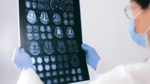
This means that the areas of hyperactivity that a person with FCD has on a brain scan likely harbor neurons with FCD-related gene mutations. Studies of resected brain tissue find that the center of seizure activity is marked by a higher percentage of cells carrying mutations [2].
These neurons may also be misshapen, similar to those observed in brain tissue from people with tuberous sclerosis complex (TSC), a genetic condition that results in tumor growth throughout the body, FCD, and seizures. This similarity gave the first clue that FCD could result from genetic mutations that affect the same processes implicated in TSC. TSC is caused by mutations to two genes, TSC1 and TSC2, which act as tumor suppressors. TSC1 and TSC2 also impact a crucial “housekeeping” pathway inside cells called the mTOR pathway, which controls cell growth, proliferation, and metabolism.
Indeed, other parts of the mTOR pathway have been implicated in FCD in the past decade, based on genetic studies of brain tissue from FCD patients. The associated genes in the mTOR pathway include the MTOR gene, P1K3CA, AKT3, TSC1, TSC2, RHEB, and DEPDC5.
But these genes still do not account for all cases of FCD. Because the mTOR pathway forms a complicated web of interacting components, other causes of FCD may lie within the mTOR pathway itself, or beyond, in yet undiscovered players. The new study by Tidball and Parent took both strategies, by looking in depth at genes belonging to the mTOR pathway, and broadly at the entire genome.
Discovery in a Dish
Drs. Andrew Tidball, Jack Parent and colleagues designed a system to explore the effects of turning off genes, one by one, to see if they resulted in a telltale sign of FCD: elevated levels of the pS6 protein, which normally helps initiate protein manufacture in a cell. In FCD, the misshapen cells that drive seizures show high levels of pS6, which is a sign of an overly active mTOR pathway.
Though this kind of experiment could take place in any kind of cell, the researchers wanted to utilize human neurons to better simulate the situation in the brain. Using stem cell techniques, the researchers grew neurons that were transformed from human skin cells in a laboratory dish, where they could be kept alive for days.
To turn genes off, the researchers introduced gene editing tools into human neurons using the Nobel prize-winning CRISPR method. This method provides a way to modify DNA in living cells. Typically, each cell took up a CRISPR molecule that switched off one gene. In all, the researchers probed the effects of over 100,000 of these molecules in over 100 million human neurons.
The researchers evaluated individual neurons for their pS6 protein outputs. Neurons with exceptionally high levels of pS6 resembled FCD cells and were therefore analyzed more carefully. This analysis confirmed some previous FCD genes, like DEPDC5, which reassured the researchers that their dish experiments could tell them something real about FCD biology.
Even more intriguing were the new genes identified. The researchers focused on six of them: HIP1, PIK3R3, LRRC4, EIF3A, TSN, URI1. Notably, two of these — HIP1 and PIK3R3 — were identified last year in studies of brain tissue from FCD patients [3], which further validates this approach.
Just as a busy intersection acts as a hub for multiple roads, the mTOR pathway gathers information from multiple cellular processes, then transforms and sends it out through other routes. Further experiments showed that the new genes influenced both the “before” and “after” of the mTOR intersection: HIP1 and PIK3R3 acted on an input to the mTOR pathway, while the other four genes seemed involved in its outputs. Thus, different disruptions to the network of interacting genes and resulting proteins surrounding mTOR may create multiple paths to FCD.
Maximal Impact
These findings could help define what researchers should look for once they obtain precious brain tissue from FCD patients to make a diagnosis. What’s more, this dish method could provide a way to test the effects of any genetic anomalies that turn up in FCD patient brain tissue. Many genetic anomalies are harmless, which means researchers need ways to sort the blameless ones from the FCD-related ones. For example, this method could be used to assess any genetic variants identified from the tiny number of brain cells that remain stuck to electrodes after they have been used to probe seizure sites in the brain while evaluating patients for surgery. Related work by CURE Epilepsy grantees Drs. Gemma Carvill and Elizabeth Gerard at Northwestern University and Dr. Alicia Goldman at Baylor College of Medicine is seeking to find a way to recover DNA from cells left on electrodes to identify the genetic causes of FCD.

Literature Cited:
- Tidball AM, Luo J, Walker JC, Takla TN, Carvill GL, Parent JM. Genome-wide CRISPRi Screen in Human iNeurons to Identify Novel Focal Cortical Dysplasia Genes bioRxiv 2023.12.13.571474.
- Lee WS, Stephenson SEM, Howell KB, Pope K, Gillies G, Wray A, et al. Second-hit DEPDC5 mutation is limited to dysmorphic neurons in cortical dysplasia type IIA Ann Clin Transl Neurol. 2019 Jul; 6:1338-1344.
- Chung C, Yang X, Bae T, Vong KI, Mittal S, Donkels C, et al. Comprehensive multi-omic profiling of somatic mutations in malformations of cortical development Nat Genet. 2023 Feb; 55: 209-220.


