Episode Overview
This week on Seizing Life® we take a look at diagnostic and surgical tools that assist physicians in localizing and removing areas of the brain that produce seizures. In an episode recorded this past November at Epilepsy Awareness Day at Disneyland, Kelly speaks with several exhibitors to learn more about current and emerging technology that is helping to improve the lives of those with epilepsy.
Episode Transcript
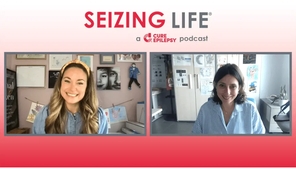
Kelly Cervantes: Hi, I’m Kelly Cervantes, and this is Seizing Life, a biweekly podcast produced by CURE Epilepsy.
Today on Seizing Life, we present interviews from our time at Epilepsy Awareness Day at Disneyland this past November. We spoke with several exhibitors about current diagnostic and surgical technology that can make a big difference in localizing and operating on areas of the brain where seizure activity is occurring.
First, we spoke with Kristen Kotsimbos of Medtronic about the Stealth Autoguide, which assists physicians in placing electrodes in a patient’s skull to localize seizure activity.
Kristen, thank you so much for chatting with us today. This is the Stealth Autoguide.
Kristen Kotsimbos: Correct.
Kelly Cervantes: What is it? And how do doctors use it?
Kristen Kotsimbos: Well, thank you for asking. I’m Kristen Kotsimbos with Medtronic. Stealth Autoguide is actually used in conjunction with self-navigation. So we don’t have that here today, but what you’ll see here is there’s a monitor, and then there’s a camera here. And what it is, it’s like GPS-type technology or navigation for brain and spine surgery. In this case, for the brain.
So, what we do is we take an MRI or CT of the brain, load it into this computer system, and then trace the contours of the face to match it up with the actual patient. We would have an instrument that just traces along these contours, and then you’d be able to see when you put instruments into the brain where you’re at on a map. So, it’d be like navigation or GPS.
And so, these spheres here, they would be reflecting off of the camera right here. And that would be the general concept of navigation, where the cranial robot comes in, which is autoguide, is to place with precision and accuracy of less than a millimeter leads. So, electrode lead placements or for RNS, SEEG and/or for Visualase, which is laser ablation. So we can do that in as small as a 2.4 millimeter burr hole. And so, it’s a very fast recovery. It’s something that you could go home the next day.
Kelly Cervantes: Oh, wow.
Kristen Kotsimbos: And so, it’s minimally invasive surgery with a precise trajectory to get really to a target with that precision of less than a millimeter.
Kelly Cervantes: So, the point of this, when a doctor might use this on a patient is going to be when they’re trying to localize where seizures are occurring. Is that correct?
Kristen Kotsimbos: That’s correct, yes. And so, they want to do it in a precise way. And so, during surgery, the actual surgeon will control the robot with this here. They’ll go ahead and get the arm into place, and then do the fine-tuning with this controller to get directly to that target.
And we also have another piece of equipment called the O-arm. In my accounts, what they do is after they place the lead, they will take the O-arm, which is another imaging device that we carry. And it looks like that’s right here. It’s a CT-like-
Kelly Cervantes: Oh, wow.
Kristen Kotsimbos: … device. And what he’ll do is he’s made a plan beforehand of exactly where he wants to target.
Kelly Cervantes: Or she.
Kristen Kotsimbos: Or she. Thank you very much. He or she because we have amazing female surgeons. Once they go ahead and place that and have done all their plans, they’ll take this image and put it back onto the Stealth Navigation System, put it onto the screen, and overlay it on top to see are we in the exact area before we leave the operating room and before we do any ablation or stimulation or anything else?
So, it’s another way to double-check that they’re exactly within the parameters that they wanted to be and on target before they do treatment.
Kelly Cervantes: Now, what are the risks that a patient would want to be aware of with this type of equipment?
Kristen Kotsimbos: In this case, because we have various steps to make sure that we’re accurate before we do treatment, it is much less risky than doing an open craniotomy to do this.
Kelly Cervantes: The hole is so small.
Kristen Kotsimbos: Correct. And so less chances of just bleeding and different things and complications with a larger incision. And so, there are always risks associated with surgery, but this takes it into completely minimally invasive and really lowers that risk factors much as we can for surgery.
Kelly Cervantes: I mean, talking about doing a type of brain surgery and then leaving the next day is, I mean, that’s just amazing to think about-
Kristen Kotsimbos: And to actually-
Kelly Cervantes: … that that type of thing is possible.
Kristen Kotsimbos: Right. And to get confirmation while you’re in the operating room so that you don’t have to come back and do a revision surgery or anything like that. So, all of these tools are just a great way to take all three technologies. The Stealth Navigation System, the cranial robot autoguide, and the O-arm for confirmation altogether to give you the best procedure and the best outcome.
Kelly Cervantes: That’s amazing. Thank you so much-
Kristen Kotsimbos: Of course.
Kelly Cervantes: … for explaining all of this to us.
Kristen Kotsimbos: Yes, of course. Thanks, Kelly. Yeah.
Kelly Cervantes: Next, we spoke with Scott Strong of Medtronic regarding the Visualase system, which can work in cooperation with the Stealth Autoguide to help surgeons perform laser ablation surgery.
Hi, Scott.
Scott Strong: Hi, there.
Kelly Cervantes: Thank you so much for chatting with us today.
Scott Strong: My pleasure.
Kelly Cervantes: All right. So, we are discussing the Visualase equipment.
Scott Strong: Correct.
Kelly Cervantes: What is it? And how do doctors use it?
Scott Strong: Visualase is MRI-guided laser ablation therapy.
Kelly Cervantes: Okay.
Scott Strong: Laser interstitial thermal therapy.
Kelly Cervantes: We just learned about the Stealth Autoguide. So, this is what they would use, once they have located the part of the brain where the seizures are emanating from, this is the next piece of equipment that they use.
Scott Strong: Correct. This device, it’s a catheter-based system, used to deliver a fiber optic laser to specific targets in the brain. So, you saw the auto guide earlier. Autoguide is a frameless stereotactic system for inserting different types of devices into the brain.
Stereotactically, the visual laser itself can be inserted with the auto guide. Visualase is a fiber optic laser. It’s delivered to target. It’s used for ablation of targets for epileptic foci, so for medically refractory, epileptic foci, brain tumor, radiation necrosis, those are all minimally invasive targets that we can go after with Visualase versus an open resection.
Kelly Cervantes: So, it’s just a small targeted area. Is this the same thing where the procedure is done and you get to go home the next day?
Scott Strong: Yes.
Kelly Cervantes: Or is this more involved?
Scott Strong: In many cases, because of this minimally invasive nature, patients sometimes go home the next day, so we hear a lot of patients get discharged the very next day. Versus open resection, it just requires a 3.2-millimeter drill hole, so just a tiny stitch. A purse string stitch, I think, is what they call it sometimes. In many cases, they get discharged the next day.
Kelly Cervantes: That’s incredible. And what does the future of this equipment look like? I feel like science and… It’s just moving so quickly. I mean, the fact that you can do this, and it’s such a small incision that needs to be made, you can so acutely target the part of the brain that needs to be removed. Do you know where this technology is headed from here?
Scott Strong: Even though MRI-guided laser interstitial therapy has been around for many years now, I want to say maybe almost a decade, it’s still new cutting-edge technology. We’re still in the phase where we have a lot of centers that are still adopting this versus other methods of ablations such as RF or doing open surgeries.
With the increased utilization of stereotactic SEEG, because of robotics, we see more customers moving towards using laser because they can more precisely identify the area of focus where seizures are coming from, and they can target it robotically and more minimal invasively, and we can use a laser to treat that versus open resection.
Kelly Cervantes: So, it sounds like it is the clinicians that need to catch up to the technology currently. The technology is available, and it’s just the community needs to come to it.
Scott Strong: The technology is available. It’s widely used in centers across the US. At least over 125 centers were done over far exceeding 8,000 cases to date.
Kelly Cervantes: Wow.
Scott Strong: Just here in the US alone.
Kelly Cervantes: Well, amazing, Scott. Thank you so much for teaching us about the Visualase system.
Scott Strong: My pleasure.
Brandon: Hi, this is Brandon from CURE Epilepsy. Did you know that 30% of those diagnosed with epilepsy do not respond to current medications? That is why for over 20 years, CURE Epilepsy has been dedicated to funding patient-focused research to find a cure for epilepsy. Learn more about our mission and our research by visiting www.cureepilepsy.org. Now, back to Seizing Life.
Kelly Cervantes: Gabriele Richardson spoke with us about MEG, a non-invasive imaging technique that can be used in conjunction with EEGs and MRIs to localize seizure activity more precisely in preparation for epilepsy surgery.
Gabriele, thank you so much for chatting with us today. I am so excited to be able to speak with you directly because I think this technology is incredible. Tell us about what an MEG is and why a doctor would want to use it.
Gabriele Richardson: So, it’s a neuroimaging device that allows physicians to detect and localize epileptic activity. If you are potentially an epilepsy patient and a candidate for surgery, it allows a neurologist to get more granular data around the brain activity and the brain networks. And as it assesses it in an EMG. So, it works in conjunction with MRI and any other neuroimaging modalities, and it allows for a far more granular of more specific picture of the brain.
Kelly Cervantes: I think I understand. I’m going to give it to you in layman’s terms, and you tell me if I’m correct. So, when MRI is a static image, but an MEG is more of a video of an EEG over the MRI. Is that accurate?
Gabriele Richardson: Yeah. An MRI would give you the typical brain image as such, whereas an MEG is actually granulas and spikes. It’s a lot of squiggly lines, basically.
Kelly Cervantes: Like the EEG.
Gabriele Richardson: Like the EEG. And that gets mapped with an MRI and with EEG as well. So, there’s training involved for epileptologist and positions to analyze the data. But because that data with in conjunction with MRI and other modalities will give you far more information as a patient for full patient.
Kelly Cervantes: And the idea that someone would want to use this ideally to try and figure out, if the seizures are localized in a certain part of the brain. So that is more data to potentially try and do a surgery, for example.
Gabriele Richardson: I mean, if you are a surgery candidate, as a patient, I would always want to have as much data as possible. So, more data will give you more information, right?
Kelly Cervantes: Yeah.
Gabriele Richardson: So, before someone opens up anyone’s brain, they should make sure they have all the data available is what I always think.
Kelly Cervantes: Yeah, absolutely.
Gabriele Richardson: And that’s why this is such a vital tool.
Kelly Cervantes: MEG, it would be considered non-invasive, correct? Because you’re not having to do any surgery. It is you go in, similar to what you would experience for an MRI, and then you leave.
Gabriele Richardson: Yeah.
Kelly Cervantes: Yeah.
Gabriele Richardson: It’s non-invasive. The only thing, it is in a big shielded room. So it’s a bit like a vault if you like, big thick door that closes. But other than that, there’s no noise because it needs to be no noise allowed sort of thing or interruptions. So, that’s why they’ve got this big shielded room. But that almost means it’s very calm in there, very quiet. It’s nothing like an MRI where you are claustrophobic.
Kelly Cervantes: Right.
Gabriele Richardson: One thing, you’ve got a lot of space, and you can lie down as well if needed. So it depends on what the epileptologist suggests and what the EEG lab suggests, but it’s very, very calming and not a invasive tool whatsoever.
Kelly Cervantes: And how many of these machines are around the country? Because I know that they’re not everywhere. It’s not like an MRI where every hospital has one.
Gabriele Richardson: Yeah, we wish there was more. It is currently around 19 in the US. It’s a mix of being used for clinical and research purposes. The clinical sites, we often do videos with them and try and raise awareness now around MEG and the States, particularly. So yeah, we have about 19 sites in the States.
Kelly Cervantes: And how could someone find out where the closest one is to them?
Gabriele Richardson: On our website now, we have a page where you have an interactive map, and you can actually find out by clicking on these different cities, which institutions have an MEG. So, that is brand new, actually, so nice that you mentioned that. That is coming out now. And then, you could potentially… And a lot of these institutions take out-of-state patients as well. And if anyone ever wants to find out a little bit more information, we are happy to provide information, so just email us, and then we will give out the information that they need.
Kelly Cervantes: No, I know that my daughter had an MEG done, and we lived in Chicago at the time, and we traveled to Milwaukee to have it done.
Gabriele Richardson: Yeah. So I mean, the other case is obviously Kate Cooper that you know-
Kelly Cervantes: Yeah.
Gabriele Richardson: … who’s traveled all the way from Virginia to Texas to have an MEG. So, we have those cases. We’re finding out about this more and more now, which helps us also raise awareness around the fact that we need more of these-
Kelly Cervantes: Yes, hopefully.
Gabriele Richardson: … around the US, right?
Kelly Cervantes: Hopefully, we will get more soon.
Gabriele Richardson: Yeah.
Kelly Cervantes: Amazing. Gabriele, thank you so much-
Gabriele Richardson: You’re welcome.
Kelly Cervantes: … for chatting with us today.
Gabriele Richardson: Nice to meet you.
Kelly Cervantes: Nice to meet you.
Ryan Chu of Zimmer Biomet explained how ROSA, the robot, assists surgeons in targeting epileptic activity deep inside the brain.
Thank you so much for chatting with us today and teaching us about ROSA. So, tell us what ROSA is and why a doctor might use it.
Ryan Chu: Yeah, absolutely. We are so glad to be here. ROSA is the robotic surgical assistant. She is a tool for brain surgeries.
Kelly Cervantes: Oh, I see what you did there.
Ryan Chu: Yeah.
Kelly Cervantes: Robotic Surgical Assistant, ROSA.
Ryan Chu: R-O Robotic-
Kelly Cervantes: Okay, yup. Uh-huh.
Ryan Chu: … Surgical Assistant, yeah.
Kelly Cervantes: I dig it.
Ryan Chu: She’s a little lady.
Kelly Cervantes: Yes, obviously.
Ryan Chu: She is a robot that helps surgeons target specific parts deep inside the brain.
Kelly Cervantes: Okay.
Ryan Chu: So, you can think of ROSA as a GPS for the brain, and a patient’s MRI or CT images are the maps for that GPS.
Kelly Cervantes: Okay. So, this is the equipment. Is this a laser equipment? How was this used?
Ryan Chu: ROSA does a lot of things. Our bread and butter is epilepsy surgery. Surgeons will use ROSA to implant what we call electrodes into patient brains, and those electrodes will monitor seizure activity and tell us where seizures are coming from. And once we know that, we can use ROSA for a therapeutic procedure.
And so, we may do a laser ablation where we stick a laser fiber into the brain and burn away the bad parts of the brain that epilepsy is coming from. Or we can use ROSA to put in electrodes for RNS or DBS, some sort of stimulation that will detect the onset of seizures and provide electrical current to prevent them from happening.
Kelly Cervantes: So, ROSA is very versatile and wears lots of hats?
Ryan Chu: Exactly. ROSA will help us diagnose epilepsy as well as treat it.
Kelly Cervantes: And how many ROSAs exist out there in the United States, specifically? Is this a pretty common piece of equipment at this point?
Ryan Chu: Yeah. At this point, we have about 140-
Kelly Cervantes: Oh, wow.
Ryan Chu: … of this across the country. I would say ROSA is probably the gold standard for the types of procedures that we’re doing with them.
Kelly Cervantes: And how invasive? I mean, you’re still talking about incisions in the skull.
Ryan Chu: Yeah, we are still making incisions, but the idea is that ROSA is a minimally invasive system. As you can see from here, this is an example patient, but there are all these colored lines going into the brain. Each one of these will represent an implant that will be going into the brain, and the idea is that we do a small quick drill hole for each of these implants.
Now, the reason it’s minimally invasive is because we’re barely cutting into the patient. We’re doing a small hole as opposed to, in the past, where if we had to access the brain, we would have to cut out large chunks of the skull.
Kelly Cervantes: Right. And how is this equipment different from other minimally invasive equipment on the market, such as the Visualase, which is one aisle over here?
Ryan Chu: Yeah. ROSA is, I think, really known for its accuracy. When we target specific centers in the brain, we’re looking at less than a millimeter accuracy, so it’s really important when we want to access those deep, really critical structures that we’re not missing, we’re not hitting any vessels or veins along the way, and ROSA helps us do that.
Kelly Cervantes: So, ROSA’s specialty is going to be those deep brain seizures that are difficult-
Ryan Chu: Exactly.
Kelly Cervantes: … to see, to access, and to treat?
Ryan Chu: Yup.
Kelly Cervantes: That is amazing. So, what does the future look like for this equipment? Are they constantly refining it? Is it advancing? And how so?
Ryan Chu: Yeah, we have a new generation in the ROSA that we’re working on right now. And again, the idea is to expand utilization to all sorts of applications. Right now, ROSA is a little more niche, it’s a little more specialized. We currently use ROSA for epilepsy surgery. We do movement disorders like Parkinson’s disease.
We’ll treat tumors and cancers with ROSA, but we want to be able to just make the system more accessible to a larger variety of surgeons and surgeon types. So the idea, again, is to continue making a system that is minimally invasive, quick, accurate, and easy to use.
Kelly Cervantes: Amazing.
Ryan Chu: Yeah.
Kelly Cervantes: Ryan, thank you so much for teaching us-
Ryan Chu: Thank you.
Kelly Cervantes: … all about ROSA today.
Ryan Chu: I’m happy to help.
Kelly Cervantes: Thank you to all who took the time to speak with us at Epilepsy Awareness Day at Disneyland. Every year, I’m inspired by the amazing, supportive, and determined community of people who attend this event. CURE Epilepsy has been proud to be a member and a leader in the epilepsy community for 25 years. We are dedicated to funding patient-focused research that will lead us to a cure for epilepsy.
If you would like to support this research, please visit www.cureepilepsy.org/donate. CURE epilepsy, inspiring hope and delivering impact.
Legal Disclaimer: The opinions expressed in this podcast do not necessarily reflect the views of CURE Epilepsy. The information contained herein is provided for general information only and does not offer medical advice or recommendations. Individuals should not rely on this information as a substitute for consultations with qualified healthcare professionals who are familiar with individual medical conditions and needs.
CURE Epilepsy strongly recommends that care and treatment decisions related to epilepsy and any other medical conditions be made in consultation with a patient’s physician or other qualified healthcare professionals who are familiar with the individual-specific health situation.


