Brain Imaging in New SUDEP Model Reveals Map of Silenced Neural Activity After Seizure
February 8, 2019
Featuring the work of CURE Grantee Dr. Stuart Cain
In a paper published in the journal Brain, researchers have identified key aspects of fatal and non-fatal seizures. Building on CURE Epilepsy- and BC Epilepsy Society-funded research that Dr. Stuart Cain and Prof. Terrance Snutch began in 2014, the team has now identified regions of the brain that become inactive after seizures in mouse models.
Depolarization of neurons is part of the process by which signals in the brain are normally transmitted between nerve cells. However, following seizures or traumatic brain injury, a severe and long lasting “spreading depolarization” occurs which instead silences activity as it moves through specific brain regions. This new research confirms that if spreading depolarization engages the brainstem, the result is fatal.
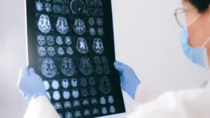
Seizures and migraine can engage similar processes in the brain, and so the team took a model with a genetic mutation found in humans causing chronic migraines and severe seizures, then monitored brain cell swelling that occurs simultaneously with spreading depolarization in real time via diffusion-weighted magnetic resonance imaging (MRI). What they found was that during fatal seizure events, depolarization spread to the brainstem, first arresting breathing; cardiac arrest followed, leading to death within a minute of the seizure. The brainstem plays an important role in regulating cardiovascular and respiratory function. In non-fatal seizures, depolarization did not spread to the brainstem.


