
Episode Overview
An EEG, or an electroencephalogram, is an essential tool in diagnosing epilepsy. And the data EEG tests produce can go beyond diagnosis by providing vital information that impacts patient treatment decisions and may help researchers unlock some of the mysteries of epilepsy and point the way towards improved treatments.
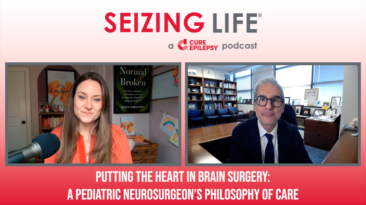
On this episode of Seizing Life, CURE Post-Traumatic Epilepsy Initiative Grantee Dr. Jeffrey Loeb from the University of Illinois at Chicago explains how EEG data is used in diagnosing, treating, and researching epilepsy.
Check out our 2020 update on Dr. Loeb’s project and the work of our PTE initiative!
Episode Transcript
Kelly Cervantes: Hi, I’m Kelly Cervantes, and this is Seizing Life, a weekly podcast produced by Citizens United for Research in Epilepsy, CURE.
Kelly Cervantes: Today I’m happy to welcome Dr. Jeffrey Loeb to the podcast. Dr. Loeb is the John S. Garvin chair, professor and head of the department of neurology and rehabilitation at the University of Illinois at Chicago. He specializes in the evaluation and treatment of epilepsy and is an expert in measuring and assessing electrical activity in the brain using EEG. He is also the recent recipient of a CURE grant in partnership with the Department of Defense for research and post-traumatic epilepsy.
Kelly Cervantes: Dr. Loeb is here today to provide a better understanding of EEG testing, the future of EEG testing, and to explain how he and his team are utilizing EEG data in his current research.
Kelly Cervantes: Dr. Loeb, thank you so much for coming in and chatting with us today about EEGs and how they work and what you’re recording. And let’s just sort of dive right into the basics. What is an EEG? What does it stand for? And what is it measuring?
Dr. Jeffrey Loeb: So an EEG is an electroencephalogram. And when you think of the brain, the brain is a bunch of wires. It’s an electrical organ. And it generates electrical energy, electrical waves. One of the problems is they’re very low amplitude. They’re very small. When you think of your heart, which also produces an electrical signal, and we put electrodes over your chest to measure your EKG or electrocardiogram, that’s a big muscle that’s going back and forth. But the brain is all these tiny little neurons. So one of the major challenges is the squiggles, amplifying them enough so that we can see and understand what they are.
Dr. Jeffrey Loeb: People have been studying brainwaves for many years, and now have atlases of all the different patterns of brainwaves that correspond to different things, when you wake, when you sleep, when you’re drowsy, when you’re having a seizure, when you’re not having a seizure, when you’ve had too much to drink. We can actually have patterns that we can see and interpret all those little squiggles.
Kelly Cervantes: So a doctor decides that a patient needs to have an EEG ordered. What is a doctor looking for in that EEG?
Dr. Jeffrey Loeb: What the EEG measures is very different than what other tests, like CAT scans or MRIs measure. In fact, when you have epileptic activity in your brain, you can’t see it with any imaging methods such as a CAT scan or MRI. It’s an electrical impulse. That electrical impulse can only be seen by wires that can measure that electrical impulse. Now these wires have to be placed on the skin closest to the brain, and those are the areas that show that abnormality. Without that test, you don’t know if there still is activity that could be consistent with seizures. You cannot tell that from an MRI or a CAT scan.
Kelly Cervantes: So the EEG is the only test that we currently have in science, aside from just sort of the naked eye and seeing someone have, but to physically measure a seizure?
Dr. Jeffrey Loeb: Absolutely.
Kelly Cervantes: Which speaks to how important this test is. So a patient goes into the hospital, and they are connected, and then sort of talk to us about there’s a couple of different types of EEGs that can be done.
Dr. Jeffrey Loeb: We do it in a very standardized way. So we put electrodes at distinct locations on the scalp, so every patient has the same arrangement, and that way we can go back to our atlases and see what it should be and what it shouldn’t be based on experience. Most of the time we do EEG, we put electrodes on the scalp with various adhesives to make them stick, sometimes stronger ones when we do longer term EEGs. One of the important things-
Kelly Cervantes: I’m going to stop you there for just a second. So what would you consider a short term EEG versus a longterm EEG? And when would a doctor order one versus the other?
Dr. Jeffrey Loeb: So we use the term routine EEG versus longterm video EEG. So a routine EEG is often anywhere from 20 minutes to an hour. The point I was going to make is that you only see what’s going on during that window that you’re looking. So if there happens to be an electrical discharge within that short 20 minute period, you may capture it. If it happens five minutes after you disconnect the wires, you miss it. And that’s why we do longer term studies because you increase the odds of capturing some of those abnormal brainwaves that are seen in people with epilepsy.
Kelly Cervantes: So I’m laughing because I don’t know how many doctors and patients and parents that I’ve spoken to that you hook the… They have a seizure at the exact same time every day, but then you hook them up to an EEG, and-
Dr. Jeffrey Loeb: When you go to the doctor, it’s gone. Right?
Kelly Cervantes: Right. And then they don’t have it, which makes this that much more complicated to diagnose and to treat and to locate when you only have one method for measuring and identifying.
Dr. Jeffrey Loeb: Well, sometime we get tricky or nasty, and we try to induce you to have your seizures while we’re recording. So during a routine EEG, we may flash lights. We may ask you to hyperventilate, breathing really hard until you get a little woozy. And those are things that can actually induce some of those abnormal wave forms.
Dr. Jeffrey Loeb: During the longterm studies, we’ll make you stay up all night, all the things that you’re told not to do because you’ll have a seizure. We’ll stop your medications. We’ll make you stay up all night. We’ll say, well, I only get seizures when I get stressed. So we bring that ex-boyfriend in there at some point and say, now there’s stress. We want to see the seizures while the EEG is on so we can characterize them, localize them, and make sure we’re doing the best treatment.
Kelly Cervantes: So the EEG is completed. You have your data. How are you reading that data? How do you use that to then help treat the patient?
Dr. Jeffrey Loeb: So we sit in rooms with computer screens, and we click page by page by page by page. So each page may have about 15 seconds of the brainwaves. If you are there for 24 hours, or four days, there’s a lot. Sometimes we click the pages quickly. We have various ways of changing the montage, which is how the electrodes talk to each other. Every electrode, to see an electrical signal, you have to have a positive lead and a negative lead. And if you orient that in a different way, you can see different directions of electrical activity. So we can modulate that montage. We can play with it. We can change the amplitude.
Dr. Jeffrey Loeb: Now EEGs are all digital. In the old days they were printed by these little, I don’t know if you’ve seen the lie detector tests with the little pen going back and forth. Those were how we did EEG. It would have like 20 pens and big, big sheets of paper. And it was actually kind of fun because you have these giant pads of paper, and you’d be flipping these pages. And every time you’d flip a page, it’d be like another 20 seconds of EEG. And you get really good. And it’s kind of a gratifying feeling to flip those pages. I kind of miss it.
Dr. Jeffrey Loeb: But yeah, we have, again, references. We have people who are experienced electroencephalographers who read the EEGs, who have a lot of experience seeing the different wave forms and patterns. And then we draw up a report that goes back to the doctor and says, this is our opinion of what we saw.
Dr. Jeffrey Loeb: If we’re lucky, we capture the epileptic discharges and/or seizures that allow us to localize where they’re coming from. The closer you are to an electrode that produces that signal, the closer in the brain that is. And because we put the electrodes all across the brain, say it’s coming from your right temporal lobe, we’ll see the electrodes on the right temporal area have a lot more amplitude of the signal than, say, the left side or other parts of the brain. And that’s how we localize where the seizures are coming from.
Brandon: Hi, this is Brandon from Citizens United for Research in Epilepsy, or CURE. Since 1998, CUre has raised more than $70 million to help fund more than 235 research grants in 15 countries around the world. Learn more at www.cureepilepsy.org. Now back to this episode of Seizing Life.
Kelly Cervantes: What is the difference between when a doctor says that they see epileptiform activity versus a spike versus a seizure?
Dr. Jeffrey Loeb: I mean, it’s kind of a funny question because even as doctors who think, or at least come across to our patients that we know everything about everything, we don’t. And if you take something that looks like a spike on an EEG, and you show it to 10 different doctors, you will get some disagreement. So sometimes it’s important to get another opinion, even with the same study, or again, change the electrical pattern, the montage, so that you can see it differently. Sometimes you have to repeat it or do it again later because there’s a lot of gray scale in reading EEGs. They’re squiggly lines. And sometimes the squiggly lines match the textbook. And sometimes they’re just not quite there. Sometimes they’re different.
Dr. Jeffrey Loeb: So another thing that’s really important is seeing the same thing over and over again. If there’s a certain part of the brain that’s epileptic, you’ll see it now. A minute later on the EEG, you’ll see it again. Five minutes later, you’ll see it. And again, when you start looking at these EEGs, you realize that people with epilepsy don’t just have seizures. They have these abnormal epileptic discharges often all the time.
Dr. Jeffrey Loeb: Another important point is the limitations, as we talked about this low signal that’s being generated from the human brain compared to say the heart. And we probably miss a lot of things that might be deeper and not on the surface. And in those cases, particularly in the cases we’re thinking of surgery, we’ll put intercranial electrodes. And we’ll do surgery, remove the skull, put electrodes directly on the brain surface, and you can see you about tenfold more electrical activity, things that you completely miss on the surface EEG.
Kelly Cervantes: Wow.
Dr. Jeffrey Loeb: The other thing we do is we never do EEG by itself. We will do an MRI. We will do PET studies. We will do other studies that corroborate what-
Kelly Cervantes: What is a PET study?
Dr. Jeffrey Loeb: Positron emission tomography. So one thing we do is look at the metabolism of the brain. So you inject it with glucose, which is food for the brain. And if its uptake goes higher or lower in some areas of the brain, you see that on the PET scan. And that can correlate with an area that has those epileptic waveforms. And when things are concordant, when you see the PET scan abnormality, when you see something on the MRI, like sclerosis or dysplasia, that is the same location where you see the electrical signals, then you say, aha, we’ve got it. And we can go after it and do surgery and help that person.
Kelly Cervantes: So combination, with the EEG, with the MRI, with potentially putting the electrodes directly on the brain, one of the ultimate end goals here is to try and localize where those seizures are coming from.
Dr. Jeffrey Loeb: So for surgical workup, that’s what we do. But we also use it just diagnostically. Sometimes people have a funny spell, may happen over and over again. I get this weird feeling, I get scared for no reason. And we don’t know whether it’s a seizure or not. Anything that you can do or feel normally can be a seizure based on where in the brain that seizure occurs. So the EEG, when we do in the hospital or at home with a continuous EEG and video going 24/7 allows us to see those spells and see if we see epileptic activity associated with them or not. Sometimes people have seizures, and they’re totally unaware they’re having seizures. So we see the seizure, and the person’s completely unaware they just had one. Other times somebody says, I just had a seizure, I just had my spell. And we see nothing on the EEG. Sometimes you get both that go together.
Dr. Jeffrey Loeb: And in some cases they’re not seizures. So one possibility is, yes, the focus is too deep. We can’t see it. And we have to think about doing the intercranial recordings. Another possibility is, well, maybe it’s just not a seizure. Maybe you’re having anxiety or getting scared for other reasons. And we can stop people from being put on medications they don’t need to be put on, getting treatments they don’t need. So it’s really very important in diagnosis. And that’s why the longterm studies are so important because if you capture the spell, you’ve got the best chance to see if it’s a seizure or not or if it’s something else.
Dr. Jeffrey Loeb: And you can also see if there are other epileptic activities between seizures, which are very common in people with epilepsy. You have these what are called interictal, which means between seizure spikes, discharges, slowing, other areas that we see abnormalities, that diagnostically help us determine that that person does indeed have epilepsy and how severe it is.
Kelly Cervantes: And correct me if I’m wrong, but you can also then look at that and diagnose a particular form of epilepsy.
Dr. Jeffrey Loeb: Correct.
Kelly Cervantes: So if you see the hypsarrhythmia pattern, you can diagnose infantile spasms. What other syndromes can you diagnose. Or types of epilepsy can you diagnose directly?
Dr. Jeffrey Loeb: There’s Lennox-Gastaut. There’s primary generalized epilepsy, juvenile myoclonic epilepsy, temporal lobe epilepsy, frontal lobe epilepsy, all those different kinds. The EEG is really one of the best indicators of that type. And then there’s some medications that work better, as we know, for some kinds of epilepsy and not others. So by having the EEG pinpoints the type of treatment that you should have.
Kelly Cervantes: Where are you seeing the future of EEGs? Where is that technology headed? And what can people look forward to?
Dr. Jeffrey Loeb: So I think we have to try to understand what is epilepsy, and particularly focal forms of epilepsy. In other words, starting in one spot of the brain or another or multiple spots in the brain. And then if you think of the brain being a bunch of wires, like a circuit board, you think of something called a network. So the normal network in your brain allows you to talk, to feel, to move. But these networks are abnormal because they cause these spontaneous discharges and symptoms that come out of nowhere.
Dr. Jeffrey Loeb: So what we really need to do is define these networks better. And we need better tools to define the networks. We’re actually working on a project where we’re taking information from the intercranial EEG, sometimes a hundred electrodes on the surface of the brain, and using really fun math to figure out what these networks look like. And then we test it on the next day and the next day. We keep seeing the same over and over again. So working with our engineering colleagues, we’ve discovered that each person has their own special network that we can then map. And it goes on between the seizures. It’s always there.
Dr. Jeffrey Loeb: And once we discover this, maybe we can do a better job of fixing the networks, of rewiring the networks, either with drugs, stem cells, treatments, lasers, noninvasive approaches, but you have to understand it. Otherwise we’re kind of shooting in the dark. So I think that’s a real important advance that’s coming along.
Dr. Jeffrey Loeb: The other thing is can we see these epileptic areas without putting electrodes directly on the brain surface? Wouldn’t it be great if we had a machine, a box, an MRI machine type thing where you could go in there and just do a scan, and here’s your network, and here’s the treatment. The next day you walk out. You don’t have seizures anymore. That would be amazing.
Dr. Jeffrey Loeb: And it’s not so much just the seizures, as we all know, and I’m sure you know from personal experience, it’s more than just about the seizures. It’s about what’s going on in the brain, how these abnormal networks are not only involved in seizure generation, but in how you feel, how you behave, how your development as a child.
Kelly Cervantes: Very few people with epilepsy are just dealing with seizures.
Dr. Jeffrey Loeb: And we need to think more about what goes on between the seizures, not just the seizures itself.
Kelly Cervantes: Absolutely. So speaking of research, and you are a recipient of one of CURE’s grants for our special project in conjunction with the Department of Defense in post-traumatic epilepsy. Can you talk with us about that research and what you’re looking at?
Dr. Jeffrey Loeb: I’m very excited about this project. People who get brain injuries, a large portion get epilepsy. And we don’t know why, and we don’t know how to stop it. So one of the things that happens when you have a impact to the brain is blood, just like you cut yourself, you get a little bleeding. You hit your head hard, you get bleeding in the brain or around the brain. Now blood is something that causes pain. You feel pain. When it’s in the brain, it causes excitability. It causes epileptic discharges. So our study is to focus on the role of blood surrounding the brain and how that changes the brain to produce it to have seizures.
Dr. Jeffrey Loeb: And there’s a group of patients who come in. We have a very busy intensive care unit of people who bleed around and in their bran. So we’re going to try to take advantage of big data, artificial intelligence, and take these entire hospital courses of very sick patients, some who develop epilepsy, and some that don’t. Look at every CAT scan, every EEG. Turn these into numbers, look at every medicine they take and build a dashboard for hundreds of patients.
Dr. Jeffrey Loeb: Then we’re going to go back and say, in this longitudinal data series, what predicts who gets epilepsy and who doesn’t? And what should we be doing in those weeks in the ICU to prevent people from getting epilepsy six months, a year later?
Dr. Jeffrey Loeb: Now in parallel with this project in our patients, we do experiments in rats where we also induce the bleeding around the brain. And then we put little EEGs on our rats, and we record their brain waves for months and watch how they progress to form seizures. And then with the animal studies, we can do further molecular tests, tests on tissue that we can’t do in people with epilepsy.
Dr. Jeffrey Loeb: So we put this all together, and the third part of our project is to look at the rat EEGs, look at the human EEGs, and build a really cool database that links everything together so we can go back and forth between what we see in the animals and what we see in humans so that if we develop a drug and treat that animal model, it’ll actually go back and work in humans. So it’s an amazing project. A lot of my colleagues, Dr. Fernando [inaudible] is head of our stroke group as part of this. Dr. [Inaudible], our stroke epidemiologist, is part of this. We have amazing students, MD-PhD students, working on the models and so forth. And CURE has helped us bring all these really smart people together to pioneer this. And once we understand how blood causes epilepsy, we’re going to understand how a lot of things cause epilepsy.
Kelly Cervantes: Absolutely. It is so exciting to me to hear about the progress being made because this will help patients, and it will help families. And hopefully it excites future researchers and future doctors to enter the field because, goodness knows, we need all the help we can get.
Kelly Cervantes: Dr. Loeb, thank you so much for your continued hard work, for coming on the podcast today, but most importantly for your dedication to this field and to the patients. And we are all just so grateful for everything that you do for this community. So thank you so much.
Dr. Jeffrey Loeb: Thank you for having me.
Kelly Cervantes: Thank you again, Dr. Loeb for sharing your expertise on EEGs and discussing how EEG data is being utilized in your current research. Research like that conducted by Dr. Loeb and his team is what will lead us to a better understanding of epilepsy and more effective treatments for patients. That’s why CURE is partnered with the Department of Defense to fund patient focused research into traumatic brain injury and post-traumatic epilepsy.
Kelly Cervantes: To support cure, please visit www.cureepilepsy.org/donate. Your generosity is greatly appreciated. Thank you.
Speaker: The opinions expressed in this podcast do not necessarily reflect the views of CURE. The information contained herein is provided for general information only and does not offer medical advice or recommendations. Individuals should not rely on this information as a substitute for consultations with qualified healthcare professionals who are familiar with individual medical conditions and needs. CURE strongly recommends that care and treatment decisions related to epilepsy and any other medical condition be made in consultation with a patient’s physician or other qualified healthcare professionals who are familiar with the individual’s specific health situation.
More from Seizing Life