

Brain Imaging May Help Predict Post-Traumatic Epilepsy After Brain Injury
Originally published in Epilepsia
A recent study investigated whether brain inflammation following a traumatic brain injury (TBI) could help identify animals that are more likely to later develop post-traumatic epilepsy (PTE). PTE can develop weeks, months, or even years after a brain injury, and researchers are interested in finding ways to identify those at greatest risk during the period before seizures begin.
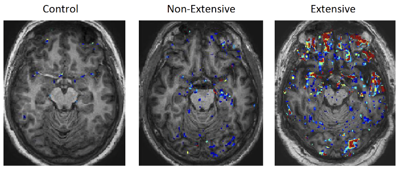
Researchers used a type of brain scan called TSPO PET imaging to measure inflammation in rats with moderate-to-severe TBI. Another group of rats that underwent a similar procedure but without brain injury served as controls. A subset of rats with TBI—18%—later developed spontaneous recurrent seizures, while none of the control animals developed seizures.
The researchers found that inflammation remained elevated in the brains of injured animals for weeks after TBI. However, the relationship between inflammation and epilepsy was more complicated than simply “more inflammation means a higher risk of epilepsy.” At four weeks after injury, rats that later developed epilepsy actually had lower TSPO signals in several brain regions than rats that did not develop epilepsy. These differences helped distinguish animals that later developed PTE from those that did not.
The injured rats also experienced problems with learning, memory, and other behaviors. The findings suggest that the location and timing of inflammation may be more important than the overall amount of inflammation. TSPO PET imaging could eventually help researchers predict who is most likely to develop PTE, and other chronic TBI-related deficits, although additional research is needed to determine whether these findings apply to people with TBI.
Related Research News