
Network Properties Revealed During Multi-Scale Calcium Imaging of Seizure Activity in Zebrafish
February 27, 2019
Understanding neuronal networks to find a critical window for therapeutic seizure control
Featuring the work of former CURE Grantee Scott Baraban
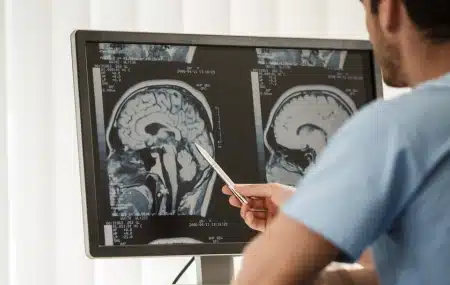
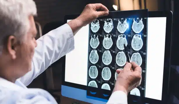
Seizures are characterized by hypersynchronization of neuronal networks. Understanding these networks could provide a critical window for therapeutic control of recurrent seizure activity i.e., epilepsy. However, imaging seizure networks has largely been limited to microcircuits in vitro or small “windows” in vivo.
Here researchers combine fast confocal imaging of GCaMP-expressing larval zebrafish with local field potential (LFP) recordings to study epileptiform events at whole-brain and single-neuron levels in vivo. Using an acute seizure model (pentylenetetrazole, PTZ), they reliably observed recurrent electrographic ictal-like events associated with generalized activation of all major brain regions and uncovered a well-preserved anterior-to-posterior seizure propagation pattern. The researchers also examined brain-wide network synchronization and spatiotemporal patterns of neuronal activity in the optic tectum microcircuit. Brain-wide and single-neuronal level analysis of PTZ- and 4-aminopyridine (4-AP)-exposed zebrafish revealed distinct network dynamics associated with seizure and non-seizure hyperexcitable states, respectively.
Neuronal ensembles, comprised of coactive neurons, were also uncovered during interictal-like periods. Taken together, these results demonstrate that macro- and micro-network calcium motifs in zebrafish may provide a greater understanding of epilepsy.
Latest from the Community